What Is FUS? Brain Targets Conditions Am I a Candidate? My Procedure Outcomes FUS vs DBS FAQ Request Consultation
Division of Functional and Epilepsy Neurosurgery
Interactive Patient Education

Focused Ultrasound
for Movement Disorders

MRI-guided focused ultrasound (MRgFUS) is an incisionless surgery that uses converging sound waves to treat tremor and other movement symptoms, with no cuts, no implants, and real-time feedback while you are awake. Explore how it works, whether you may be a candidate, and how it compares to other options.

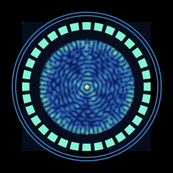
~900–1,000 ultrasound beams converge at a single point

What Is Focused Ultrasound?

MRI-guided focused ultrasound (MRgFUS, or just FUS) focuses hundreds of ultrasound beams through the intact skull to a precise target deep in the brain to create a small, controlled lesion (disconnection) that interrupts the abnormal circuit driving your symptoms. There is no incision and no implanted device.
🎯

Precisely Targeted

Real-time MRI thermometry measures the temperature at the exact focus point, allowing the care team to confirm accuracy before the definitive treatment is delivered.

🧠

No Incision

The ultrasound beams pass through the skull without cutting. There is no opening in the scalp or skull, no implanted hardware, and no general anesthesia.

🗣️

Awake & Interactive

You remain awake throughout so your care team can test symptom improvement in real time. Low-energy test pulses are given first so you can provide feedback before the permanent lesion is made.

🏥

Same-Day Discharge

Most patients are discharged the same day. Surgical recovery is typically quick and no follow-up for device programming (as for DBS) is needed.

How It Works — Step by Step

👆 Select a step to learn what happens — and why.
Important to Understand

Surgery Without an Incision

MRgFUS is surgery because it achieves its effect by creating a permanent, targeted change in brain tissue. DBS, on the other hand, requires the surgical implantation of a device to deliver reversible, adjustable stimulation. Both are effective options; they work differently and suit different patients.
Note on irreversibility: Because FUS creates a permanent lesion, it cannot be "turned off" if side effects occur after the therapeutic sonication. This is a key consideration when comparing FUS to DBS.
Key Distinction vs. DBS
FUSCreates a permanent brain lesion. No device to maintain or program. Effect is immediate and irreversible.
DBSDelivers ongoing electrical stimulation via implanted electrodes. Fully adjustable and reversible — can be turned off or modified over time.
For a detailed side-by-side comparison of FUS and DBS, see below ↓
Physics of Focusing Ultrasound
For the Curious

Why Hundreds of Beams? The Physics of Focusing Ultrasound

How does ultrasound energy pass harmlessly through the skull and converge with millimeter precision on a target deep in the brain without any incision? The answer lies in the elegant physics of constructive interference. Click to explore an interactive simulation.

Take a deep dive into the physics

Ultrasound energy is delivered by individual "transducer" elements. Each transducer element on its own would spread energy across a wide volume of tissue. The waves of energy from each transducer will add or cancel, depending on their relative phases (where the peaks and troughs of the wave line up). The key is timing: when every element is delayed by exactly the right amount, all waves converge at the target simultaneously, arriving "in phase" and summing to produce an intensity far beyond what any single element could achieve.

Drag the target to steer the focus and watch each element's phase (shown by ring color) update in real time. Adjust the sliders to explore how the number of elements and phase accuracy (affected by skull density) shape the focal spot. Note that higher element counts are especially useful to correct for phase error, such as skull distortion.

Focal power (log scale) 4,096×
Low → High intensity Phase delay
Transducer elements: 64
More elements → higher focal power (N² scaling)
Phase error (skull distortion): 0%
Random phase errors simulate skull bone distortion
Phase efficiency
100%
Focal intensity (N²)
4,096×
💡
With 64 elements firing in perfect phase, peak intensity at the focus is 4,096× (64²) greater than a single element — all concentrated in a spot millimeters wide.

Which Symptoms Can FUS Help?

Select your diagnosis, then click any symptom to see how FUS typically addresses it.

👆

Select a symptom to see how FUS typically affects it.

Brain Targets

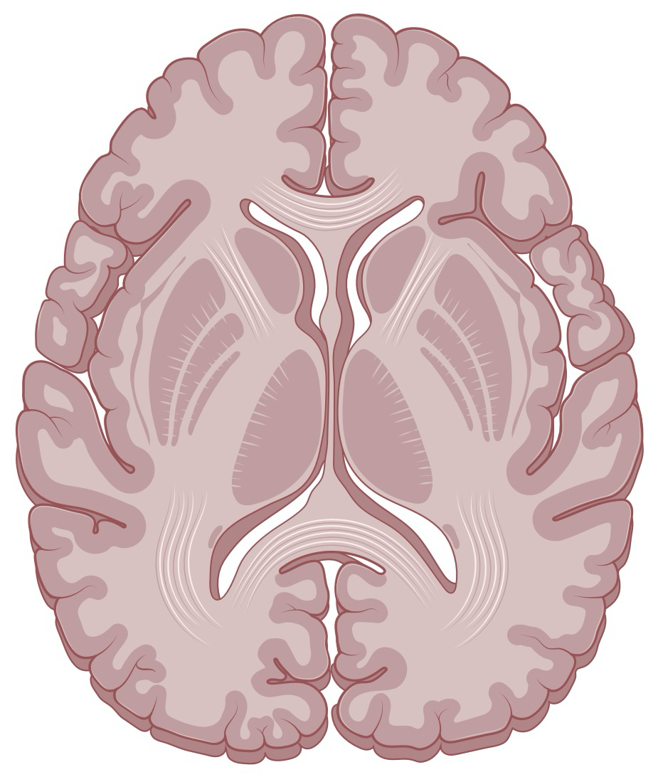
MRgFUS is FDA-approved for three brain targets, each addressing different symptoms. Select a target below to learn more.

VIM
Ventral Intermediate Nucleus
of the Thalamus
Tremor
GPi
Globus Pallidus
Internus
PD Motor
PTT
Pallidothalamic
Tract
PD Motor
Axial brain slice showing target locations VIM GPi PTT

Axial slice · Targets shown bilaterally

Unilateral vs. Bilateral Treatment

FUS creates a lesion on one side of the brain at a time. Because both sides of the brain can be affected by movement disorders, the question of whether to treat one side or both is an important part of your evaluation.

Unilateral

One-Side Treatment

A single FUS procedure treats one hemisphere of the brain and affects movement on the opposite side of the body. For most patients, the dominant hand (and thus the non-dominant hemisphere) is treated first, as this typically provides the greatest functional benefit.

Unilateral treatment is the standard first procedure. If the non-treated side causes significant disability after the first procedure, a second procedure may be considered after the required interval.

  • Addresses one side of body
  • Lower risk than bilateral
  • Can be followed by contralateral procedure if needed
Bilateral

Sequential Two-Side Treatment

Bilateral FUS is FDA-approved and is performed as two staged procedures — one per side — with a mandatory minimum interval of 9 months between treatments. This interval allows for safety assessment and full recovery of the first side.

Side 1
≥ 9 months
Side 2
Each of the two stages is a separate outpatient procedure
Why the interval? Bilateral thalamic or pallidal lesions carry a higher risk of speech and swallowing side effects. The 9-month waiting period allows clinicians to assess the full effect of the first procedure before treating the opposite hemisphere.
⚠️

Bilateral Treatment Carries Higher Risk

Treating both hemispheres increases the risk of dysarthria (slurred speech), dysphagia (swallowing difficulty), and subtle changes in cognition or balance. These risks are carefully weighed against the expected benefit, particularly for axial symptoms like voice tremor that require bilateral treatment to improve.

Am I a Candidate for FUS?

Not everyone with a movement disorder is an ideal candidate for FUS. Several factors — including diagnosis, skull anatomy, and overall health — determine whether FUS is appropriate for you.

👆

Select a category above, then tap any factor to read more.

Understanding the Skull Density Ratio (SDR)

The SDR is a CT-derived measure of how porous your skull bone is. The skull contains an inner cancellous (spongy) bone layer that absorbs and scatters ultrasound — the more porous this layer, the lower the SDR. A higher SDR indicates denser, more solid bone that transmits energy more efficiently. Drag the slider below to see how skull density appears on CT.

SDR Scale — drag to explore
0.2 0.3 (min) 0.5 0.8
Values at or above 0.3 support FUS candidacy; 0.4+ gives the most reliable results.
How It's Measured
A non-contrast CT scan of the head is sufficient. No additional radiation exposure beyond standard pre-operative imaging.
If SDR < 0.3
FUS may not be feasible. DBS has no skull density requirement and may be an alternative.
CT Appearance by Skull Density
SDR ≈ 0.40 Good candidate

What to Expect

From your first consultation through the day of treatment and beyond — here is what the FUS process looks like at each stage.

1
Initial Consultation
Weeks before
You meet with the neurosurgeon to review your history, examine your symptoms, and discuss whether FUS is appropriate. Your existing brain MRI and head CT are reviewed. If an adequate CT is not available, one will be obtained to calculate the SDR. You receive a thorough explanation of the procedure, benefits, risks, and alternatives — including DBS.
2
Multi-Disciplinary Evaluation
1–4 weeks before
A formal multi-disciplinary evaluation is completed by a team that typically includes a movement disorders neurologist and a neuropsychologist. The team assesses your suitability for FUS and/or DBS and discusses the relative merits of each for your specific situation. This evaluation may include neuropsychological testing, a movement disorders neurology visit, and a review of medications. After the evaluation, you will meet again with your neurosurgeon to discuss the team's recommendations and decide together whether and how you wish to proceed. Blood thinners may need to be paused before the procedure.
3
Procedure Day: Preparation
Morning of procedure
You arrive at the hospital having fasted overnight. Your head is shaved (the hair will regrow). A stereotactic head frame is affixed under local anesthesia. A silicone membrane and water-filled transducer helmet are placed. You are awake throughout the main procedure; frame placement is typically performed with brief, mild IV medicine for relaxation.
4
MRI Suite: Targeting & Test Sonications
~2–3 hours
Inside the MRI scanner, the treatment team uses real-time imaging to identify and confirm the brain target. Low-energy test sonications are delivered and you are asked about changes in your tremor or symptoms, and any sensations such as tingling or imbalance. The position is adjusted until the optimal focus is achieved.
5
Therapeutic Sonication
Minutes
Once the target is confirmed, the full therapeutic energy is delivered. A brief pulse raises the tissue temperature at the focus point to 55–60 °C for several seconds, creating a permanent thermal lesion approximately 3–4 mm in diameter. Many patients notice immediate improvement in motor symptoms such as tremor at this stage.
6
Recovery & Discharge
Same day
After the procedure, the frame is removed and you are observed for several hours. A brain MRI is typically obtained before discharge to assess the lesion and confirm targeting. Most patients are discharged the same day. There is no surgical wound to care for. Common temporary side effects include fatigue, mild imbalance, or tingling. Symptom benefit is typically apparent immediately.
7
Follow-Up & Monitoring
Weeks to months after
Follow-up visits assess symptom improvement, side effects, and overall recovery. A brain MRI is typically obtained at 3–6 months to confirm lesion characteristics. Medication adjustments may be made in collaboration with your movement disorders neurologist. If bilateral treatment is being considered, the decision is made at or after the 9-month follow-up.

What Can FUS Achieve?

The data below reflect published outcomes from pivotal clinical trials and real-world studies. Individual results vary; your specific outcome will depend on your diagnosis, target, and skull characteristics.

~75%
ET Tremor Reduction (VIM)
Patients with essential tremor experience an average reduction in hand tremor scores of approximately 75%, with many achieving near-complete tremor suppression.
~55–65%
PD Motor Improvement (GPi / PTT)
PD motor scores (UPDRS Part III) improve by 55–65% in off-medication state for the contralateral body, including tremor, rigidity, and bradykinesia.
~85%
ET: Meaningful Functional Benefit
The majority of ET patients report meaningful improvement in activities of daily living — including handwriting, eating, and drinking — that is sustained over years of follow-up.
5+ yrs
Durability of Effect
Long-term follow-up studies demonstrate durable tremor suppression beyond 5 years for VIM thalamotomy. A minority of patients — approximately 10–15% — experience meaningful tremor re-emergence over time. Re-treatment with a repeat FUS procedure or transition to DBS is possible for those who need it.

Common Temporary Side Effects

Temporary side effects during or immediately after FUS are common and typically resolve within days to weeks. These include: mild imbalance or unsteadiness (most common), tingling or numbness in the hand or face, fatigue, and headache. Serious complications are uncommon but include significant dysarthria (slurred speech), particularly with bilateral treatment.

FUS vs. Deep Brain Stimulation

Both FUS and DBS are effective surgical treatments for movement disorders. The right choice depends on your diagnosis, symptoms, anatomy, and personal priorities. Toggle below to compare.

FeatureFUS (MRgFUS)Deep Brain Stimulation
MechanismThermal ablation — permanent lesion in brain tissueChronic electrical stimulation via implanted electrodes
IncisionNone — no skin cutsSmall burr holes in skull + incision for pulse generator (chest)
ReversibilityPermanent — lesion cannot be undoneReversible — device can be turned off or removed
AdjustabilityNone — effect is fixed at time of treatmentFully adjustable — stimulation parameters tuned over time
AnesthesiaAwake (local); mild sedation availableVaries: awake, or asleep with robotic / MRI guidance
Hospital StaySame-day discharge (typical)2 or 3 stages, one of which requires a 1-night stay
Bilateral TreatmentStaged: two separate procedures, ≥ 9 months apartBoth sides can be treated in a single procedure
Effect OnsetImmediate (often felt during procedure)After device activation and programming (weeks to months)
Long-term MaintenanceNone — no hardware to manageBattery replacement every ~3–5 years; programming visits
MRI After ProcedureUnrestrictedConditional — head/body MRI under specific conditions

Neither FUS nor DBS is universally "better" — they are complementary tools suited to different patients and priorities. Your neurosurgeon and movement disorders neurologist will help you weigh these options based on your specific situation.

Common Questions

Is focused ultrasound really surgery if there's no incision?
Yes: we consider MRgFUS to be a form of surgery, specifically an incisionless surgery. It achieves its effect by creating a permanent, targeted change in brain tissue through thermal ablation. The absence of a skin incision is a major practical advantage, but the procedure is still a controlled, permanent intervention on the brain that requires the same careful evaluation and informed consent as any neurosurgical procedure.
Will my hair grow back?
Yes. Your head is shaved on procedure day to allow the transducer helmet to contact the scalp and transmit ultrasound efficiently. Hair regrowth is complete within a few months and is not permanent.
Does the procedure hurt?
The stereotactic head frame is placed under local anesthesia and may cause a sense of tightness or pressure that typically decreases over several minutes. During sonications, you may feel warmth, brief tingling, or head pressure; in some cases, especially for patients with low SDR, this sensation can be more intense, though brief. Some patients feel momentary dizziness during test sonications. The treatment team monitors you closely and you can communicate at any time. Most patients describe the experience as tolerable.
What if my skull density ratio is too low?
If your SDR is below 0.3, ultrasound energy cannot be delivered safely and effectively, and FUS is not a viable option for you. This is not a reflection of overall health; it is simply a characteristic of skull anatomy. DBS (deep brain stimulation) may be an excellent alternative that has no skull anatomy requirement and is effective for the same conditions.
How long does the tremor improvement last?
Long-term follow-up studies (up to 5+ years) demonstrate durable benefit for the majority of patients. However, approximately 10–15% of patients experience meaningful tremor re-emergence over time. This appears to be related to adaptive plasticity in the surrounding circuits rather than the lesion itself "growing back." If tremor does return to a disabling degree, re-treatment is possible: either through a repeat FUS procedure or, alternatively, by transitioning to DBS. The durability of GPi and PTT outcomes is still being characterized as these are newer targets.
Can I have FUS if I already have a DBS device?
Existing DBS hardware is generally a contraindication to MRgFUS, primarily because the MRI required for guidance is incompatible with active DBS systems and because metal hardware can alter the ultrasound field. Each case is evaluated individually.
How soon can I return to normal activities after FUS?
Most patients resume light daily activities within days. There is no wound to care for, no surgical recovery period in the traditional sense, and no overnight hospitalization. Driving restrictions typically apply for a short period. Most patients are back to their full routine within 1–2 weeks.

Who We Are

This educational resource was developed by the Division of Functional and Epilepsy Neurosurgery at Brown University Health to help patients and families understand focused ultrasound treatment for movement disorders. Our surgeons specialize in the full range of functional neurosurgical options — including FUS, DBS, and ablative procedures — and work closely with the movement disorders neurology team to ensure each patient receives the most appropriate, individualized care.

What Is FUS? Brain Targets Conditions Candidacy Procedure Outcomes FUS vs DBS FAQ