For one-third of people with epilepsy, two or more medications fail to stop seizures. Surgery, ranging from minimally invasive laser ablation to implantable responsive stimulators, can dramatically reduce or eliminate seizures for carefully selected patients.
Defined by failure of two or more appropriate anti-seizure medications at adequate doses, drug-resistant epilepsy (DRE) affects a substantial minority of people living with epilepsy, and surgery can help many of them.
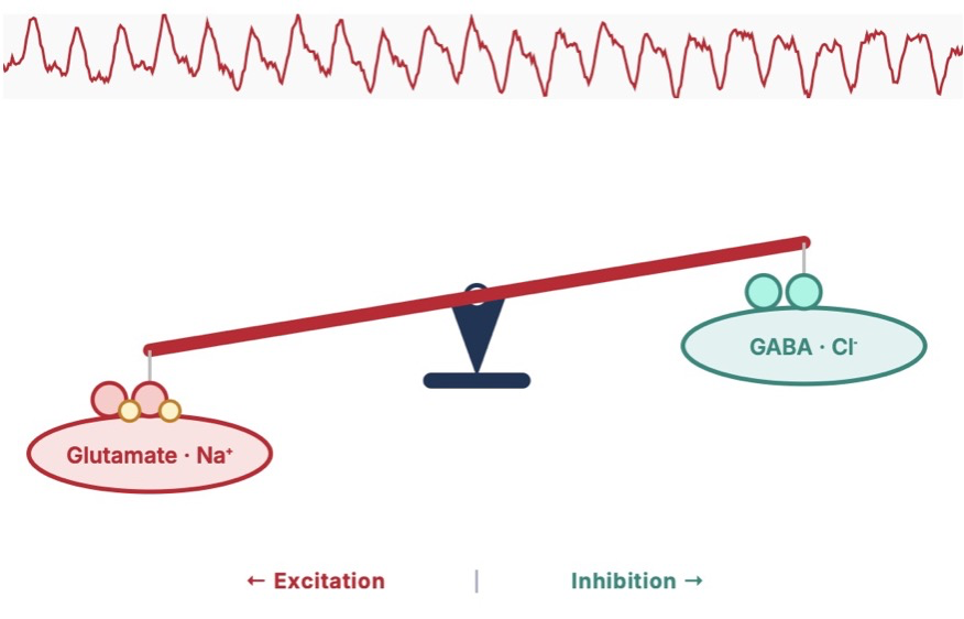
Every seizure begins with an imbalance between the brain's excitatory and inhibitory forces. Explore how this works and how treatments restore the balance.
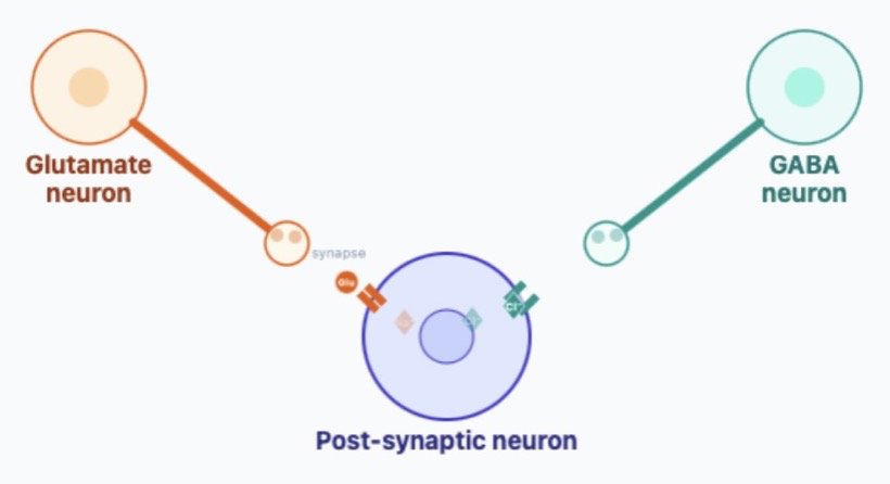
To understand what creates a seizure, it helps to see how neurons communicate. Each neuron receives signals from thousands of others in the form of neurotransmitters: these are small molecules that flow from one cell to another across a small gap (a synapse), some pushing the receiving neuron to fire (excitatory), others holding it back (inhibitory). Use the sliders to explore how these two forces compete at a single receiving ("post-synaptic") neuron. When excitation outweighs inhibition and a critical threshold is reached, the receiving neuron fires a pulse of activity (an "action potential") to influence its own downstream targets.
When medications do not fully control seizures, or when the side effects of multiple medications interfere with quality of life, surgery may be an option to reduce or eliminate seizures.
Select one or more treatments to compare. The right approach depends on your seizure type, where seizures originate, and which brain regions are involved.
Temporal lobe epilepsy (TLE) is the most frequent form of drug-resistant focal epilepsy and the most common indication for epilepsy surgery. The optimal approach depends on which structures are involved and whether one or both temporal lobes generate seizures. Select a treatment below, then click any highlighted region to learn about its anatomy and role in TLE surgery.
Understanding how seizures are classified helps you communicate with your care team. Select a category, then click any seizure type for a plain-language description.
Select a seizure type from the list to see a description
Candidacy for epilepsy surgery involves many factors. Select a category to explore what our team evaluates. Only a comprehensive multidisciplinary workup can determine whether surgery is right for you, and if so, which approach.
Select a factor to see what our team looks for during evaluation
Epilepsy surgery evaluation is methodical and sometimes lengthy, because precision matters. Click any step to expand details.
Brown University Health is designated a Level 4 Epilepsy Center by the American Epilepsy Society (AES), the highest designation, reflecting our capacity to evaluate and treat the most complex cases using the full range of diagnostic and surgical options.
Your evaluation begins with a detailed history, neurological exam, and review of all prior medications and studies. We document your seizure semiology (what happens before, during, and after a seizure), because this alone points to where seizures may be coming from.
You will be admitted to our epilepsy monitoring unit (EMU) for video-EEG monitoring to capture typical seizures on scalp electrodes, correlating the electrical signature with observed behavior.
High-resolution MRI at 3 Tesla looks for subtle structural lesions (mesial temporal sclerosis, cortical dysplasia, tumors, or vascular malformations) that may represent the seizure focus. MRI is normal in a significant proportion of DRE patients, which does not preclude surgery but adds complexity.
Additional imaging often includes FDG-PET (metabolic activity), ictal SPECT (blood flow during seizure), and magnetoencephalography (MEG). When findings from different modalities converge on the same region, confidence in localizing the focus increases substantially.
Your case is presented at our multidisciplinary epilepsy surgery conference, attended by epileptologists, neurosurgeons, neuropsychologists, neuroradiologists. The team reviews all available data and reaches a consensus on whether a seizure focus has been identified and what the next step should be.
Three outcomes are possible: (1) sufficient localization to proceed directly to surgical planning; (2) need for additional non-invasive testing; or (3) need for invasive monitoring to better define the seizure focus and network.
When scalp EEG and imaging are insufficient to localize the seizure focus, or when the suspected focus is near eloquent cortex, we proceed to invasive intracranial recording. This is a surgical procedure and requires a dedicated hospital admission, typically lasting 7 to 14 days.
SEEG (Stereo-EEG): Thin depth electrodes are placed through small skull perforations to record from deep brain structures. Our program places SEEG electrodes using a highly precise stereotactic surgical robot for image-guided placement. SEEG is particularly well-suited to mapping seizure networks that span multiple lobes or cross hemispheres.
ECoG (Electrocorticography): A grid or strip of electrodes is placed directly on the brain surface through a craniotomy. ECoG provides dense coverage of cortical surface areas and allows simultaneous cortical stimulation mapping to identify eloquent regions.
The goal of invasive monitoring is not simply to find "where seizures start" but to understand the full seizure network: how the ictal discharge propagates, which regions are necessary for seizure expression, and which adjacent areas subserve critical functions that must be preserved.
The definitive intervention is selected based on all prior findings and conference discussion. Options include open resection (craniotomy), laser interstitial thermal therapy (LITT), or implantation of a neuromodulation device (RNS, DBS, or VNS).
Laser ablation (LITT) using our Visualase or ClearPoint systems allows precise thermal destruction of the seizure focus through a single small skull opening, guided in real time by MRI thermometry. For certain targets, particularly mesial temporal structures or nodular heterotopia, LITT offers efficacy comparable to open resection with significantly less surgical exposure. Ablations can be staged or repeated to balance seizure control against functional risk.
RNS (responsive neurostimulation) is our preferred neuromodulation approach when resection or ablation is not safe or when the seizure network is too distributed for focal treatment. The RNS device continuously monitors brain activity and delivers brief stimulation pulses when it detects pre-seizure patterns, essentially closing the loop between detection and treatment. Critically, the device accumulates an objective, longitudinal record of your seizure burden that helps guide ongoing management.
Following resective or ablative surgery, most patients remain on their pre-operative anti-seizure medications for at least 6–12 months. As seizure freedom is sustained, your epileptologist will consider gradual medication reduction. The timing and pace of any taper is individualized.
For neuromodulation devices, the immediate post-implant period involves device programming and optimization, a process that continues over months as the system accumulates data from your brain. RNS programming in particular benefits from the longitudinal EEG data the device stores.
Long-term follow-up with your epileptologist and, if applicable, the neurosurgical team is essential. MRI, EEG, and neuropsychological testing may be recommended to monitor for changes over time.
For RNS patients, regular clinic visits allow the team to review stored electrocorticography data, assess detection and stimulation efficacy, and adjust device parameters. Battery longevity for RNS devices varies but is generally measured in years; battery replacement requires a minor outpatient procedure.
Driving, employment, and quality-of-life goals should be revisited at each visit. Many patients who achieve sustained seizure freedom regain their driver's license and return to activities that seizures had previously prevented.
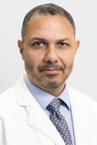
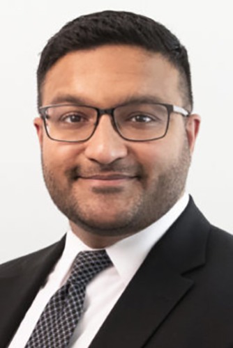
This resource was developed by the Division of Functional and Epilepsy Neurosurgery at Brown University Health to help patients and families better understand epilepsy, its causes, and the range of treatments available. Our division cares for patients with epilepsy, movement disorders, psychiatric conditions, and other disorders of neural circuit function, offering surgical and neuromodulation approaches aimed at restoring quality of life. We hope this page supports more informed and confident conversations about your care.