Drug-Resistant Epilepsy Treatments TLE in Focus Seizure Types Am I a Candidate? My Journey FAQ Request Consultation
Division of Functional and Epilepsy Neurosurgery
Interactive Patient Education

When Seizures Continue Despite Medication

For one-third of people with epilepsy, two or more medications fail to stop seizures. Surgery, ranging from minimally invasive laser ablation to implantable responsive stimulators, can dramatically reduce or eliminate seizures for carefully selected patients.

Cortical neurons

Drug-Resistant Epilepsy

Defined by failure of two or more appropriate anti-seizure medications at adequate doses, drug-resistant epilepsy (DRE) affects a substantial minority of people living with epilepsy, and surgery can help many of them.

💊
What counts as "drug-resistant"?
Failure of two appropriately chosen medications at adequate doses.
⚠️
Why uncontrolled seizures matter
Falls, SUDEP, and lost independence are real and preventable risks.
📈
How surgery has evolved
Far less invasive today, and many more patients qualify.
🕐
When to seek evaluation
Surgery is a proactive step — not a last resort.

~65M
People worldwide living with epilepsy
1 in 3
Develop drug-resistant epilepsy despite optimal medication management
>60%
Seizure freedom rates achievable in well-selected surgical candidates (temporal lobe epilepsy)
7 yrs
Average delay from diagnosis to surgical referral; evidence suggests earlier referral leads to better outcomes
Our goal as epilepsy surgeons
Not merely to reduce seizure frequency, but to determine whether seizure freedom is achievable and to work with individuals with epilepsy to find the safest, most effective path to get there.

What Is Epilepsy? The E/I Balance

Every seizure begins with an imbalance between the brain's excitatory and inhibitory forces. Explore how this works and how treatments restore the balance.

How Seizures Arise

In a healthy brain, excitatory and inhibitory activity are kept in careful balance across all regions. But a wide range of brain injuries, illnesses and developmental anomalies (including strokes, traumatic brain injury, cortical malformations, tumors, and scarring from prior infections) can disrupt this balance locally. Damaged or abnormally organized tissue may produce too much glutamatergic excitation, lose inhibitory interneurons, or alter the density and sensitivity of ion channels, tipping the local E/I ratio toward excitation. These regions of persistent imbalance lower the threshold for synchronized neuronal firing, creating a site from which seizures can originate repeatedly — what clinicians call a seizure focus. Explore below how medications can target different components of the E/I balance, and how other physiological factors alter this balance.

Brain
Neuron synapse illustration
Interactive · Cell Level
The E/I Balance on a Cellular Level: Glutamate and GABA
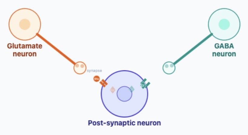
Every seizure starts at the level of individual neurons responding to a balance of excitatory and inhibitory inputs. Use the sliders to adjust the strength of excitatory (glutamate) and inhibitory (GABA) signals and watch how a neuron reacts to the opposing forces.
Explore the neuron simulation

To understand what creates a seizure, it helps to see how neurons communicate. Each neuron receives signals from thousands of others in the form of neurotransmitters: these are small molecules that flow from one cell to another across a small gap (a synapse), some pushing the receiving neuron to fire (excitatory), others holding it back (inhibitory). Use the sliders to explore how these two forces compete at a single receiving ("post-synaptic") neuron. When excitation outweighs inhibition and a critical threshold is reached, the receiving neuron fires a pulse of activity (an "action potential") to influence its own downstream targets.

Figure key
Electrical pulse
Action potential travelling down the axon
Neurotransmitter
= Glutamate (Glu)  ·  = GABA
Ion entering cell
= Na⁺ (depolarising = exciting)  ·  = Cl⁻ (hyperpolarising = inhibiting)
Transmembrane receptor
Channel opens when neurotransmitter binds
Glutamate Na⁺ influx Excitatory
GABA Cl⁻ influx Inhibitory
⚡ Glutamate
SilentHyperactive
🛡 GABA
SilentHyperactive
Try a preset:
E/I balance scale illustration
Interactive · Network Level
The E/I Balance: Medications & Triggers
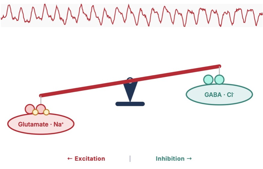
Dozens of factors — from medications to sleep to hormones — shift the balance between excitation and inhibition across entire brain networks. Explore how anti-seizure drugs restore that balance, and how common triggers tip it the wrong way.
Explore medications & seizure triggers
Excitatory / Inhibitory Balance
Live EEG Signal
Glutamate · Na⁺ GABA · Cl⁻ ← Excitation | Inhibition →
Neural Network Activity
Seizure Threshold Exceeded
How do anti-seizure medications restore the balance? Click a class to find out:
Factors that can tip the balance toward seizure. Click to explore:
Click a trigger or medication class to learn more.

Surgical Treatment Options

When medications do not fully control seizures, or when the side effects of multiple medications interfere with quality of life, surgery may be an option to reduce or eliminate seizures.

Select one or more treatments to compare. The right approach depends on your seizure type, where seizures originate, and which brain regions are involved.

🧠
Option 1
Resection / Craniotomy
Surgical removal of seizure focus
Click to learn more ›
🔆
Option 2
Laser Ablation (LITT)
Minimally Invasive Seizure Focus Ablation
Click to learn more ›
Option 3
Neuromodulation
RNS · DBS · VNS
Click to learn more ›
Measuring Surgical Outcomes: The Engel Classification
Epilepsy surgery outcomes are commonly reported using the Engel scale. Most outcome studies define success as Engel Class I, representing essentially complete seizure freedom or nearly so.
Class I
Seizure Freedom
Free of disabling seizures. May have rare auras or early post-op seizures that do not persist. The goal of resective or ablative surgery.
Class II
Nearly Seizure Free
Rare disabling seizures ("almost seizure-free"). Meaningful reduction in seizure burden with sustained benefit.
Class III
Worthwhile Improvement
Worthwhile improvement in seizure frequency or severity, but not approaching seizure freedom.
Class IV
No Worthwhile Improvement
No meaningful change or worsening in seizure burden. Guides decisions about additional or alternative interventions.

Treating the Most Common Type of Focal Epilepsy

Temporal lobe epilepsy (TLE) is the most frequent form of drug-resistant focal epilepsy and the most common indication for epilepsy surgery. The optimal approach depends on which structures are involved and whether one or both temporal lobes generate seizures. Select a treatment below, then click any highlighted region to learn about its anatomy and role in TLE surgery.

Hippocampus
Lateral Temporal Cortex
Amygdala
Perirhinal Cortex
Image source
Bollmann, Steffen, Andrew Janke, Lars Marstaller, David Reutens, Kieran O’Brien, and Markus Barth. “MP2RAGE T1-weighted average 7T model” January 1, 2017. doi:10.14264/uql.2017.266

Seizure Type Explorer

Understanding how seizures are classified helps you communicate with your care team. Select a category, then click any seizure type for a plain-language description.

Select a seizure type from the list to see a description

Am I a Surgical Candidate?

Candidacy for epilepsy surgery involves many factors. Select a category to explore what our team evaluates. Only a comprehensive multidisciplinary workup can determine whether surgery is right for you, and if so, which approach.

🔍

Select a factor to see what our team looks for during evaluation

Your Epilepsy Surgery Journey

Epilepsy surgery evaluation is methodical and sometimes lengthy, because precision matters. Click any step to expand details.

1
Phase 1: Non-Invasive
Initial Comprehensive Evaluation

Brown University Health is designated a Level 4 Epilepsy Center by the American Epilepsy Society (AES), the highest designation, reflecting our capacity to evaluate and treat the most complex cases using the full range of diagnostic and surgical options.

Your evaluation begins with a detailed history, neurological exam, and review of all prior medications and studies. We document your seizure semiology (what happens before, during, and after a seizure), because this alone points to where seizures may be coming from.

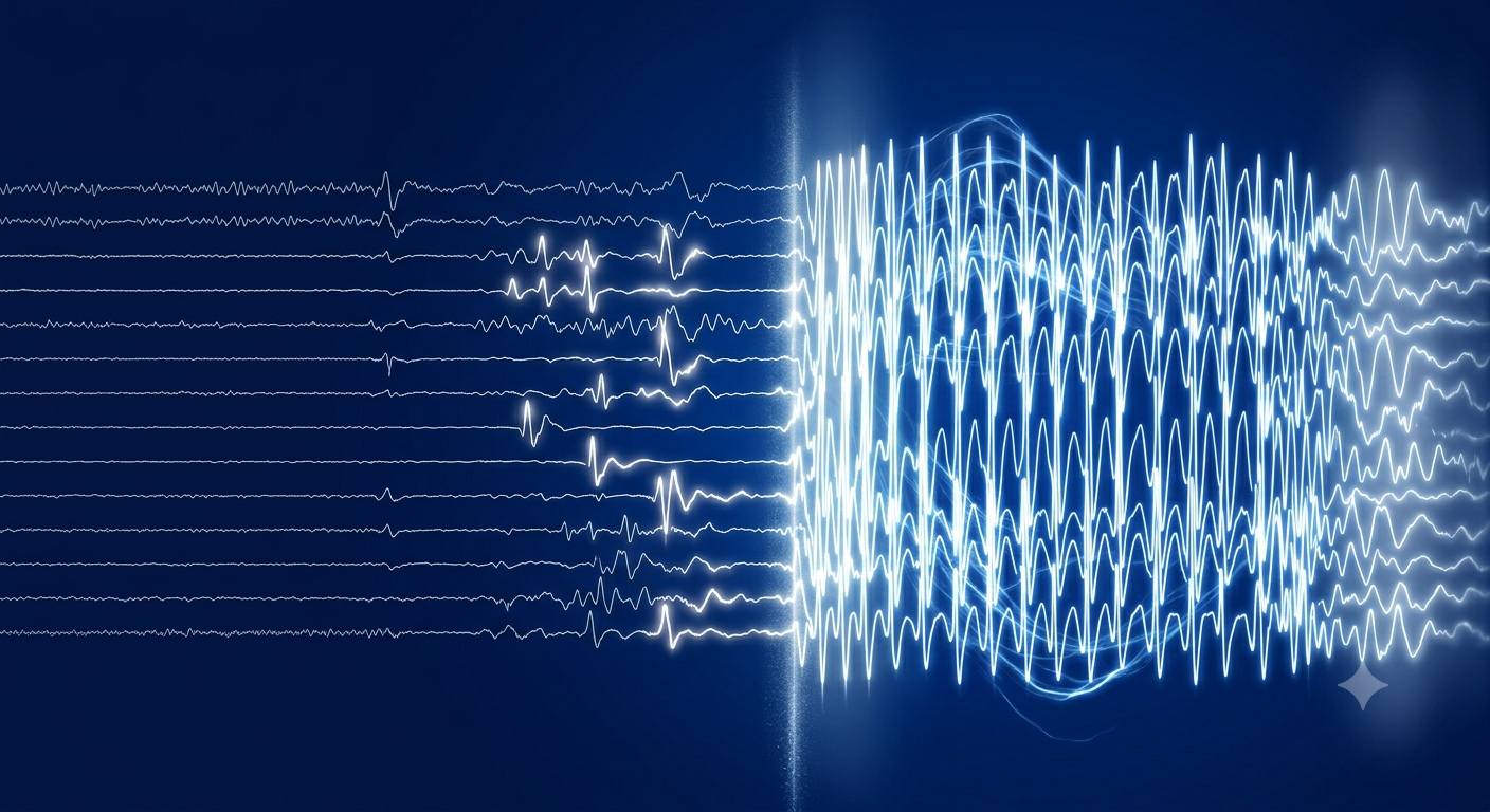
You will be admitted to our epilepsy monitoring unit (EMU) for video-EEG monitoring to capture typical seizures on scalp electrodes, correlating the electrical signature with observed behavior.

Ictal EEG recording showing seizure activity
Duration: typically 3–10 days inpatient for adequate seizure capture. Medications may be carefully reduced to allow seizures to occur safely in the monitored setting.
2
Phase 1: Non-Invasive
Advanced Neuroimaging

High-resolution MRI at 3 Tesla looks for subtle structural lesions (mesial temporal sclerosis, cortical dysplasia, tumors, or vascular malformations) that may represent the seizure focus. MRI is normal in a significant proportion of DRE patients, which does not preclude surgery but adds complexity.

Additional imaging often includes FDG-PET (metabolic activity), ictal SPECT (blood flow during seizure), and magnetoencephalography (MEG). When findings from different modalities converge on the same region, confidence in localizing the focus increases substantially.

3
Multidisciplinary Conference
Epilepsy Surgery Conference

Your case is presented at our multidisciplinary epilepsy surgery conference, attended by epileptologists, neurosurgeons, neuropsychologists, neuroradiologists. The team reviews all available data and reaches a consensus on whether a seizure focus has been identified and what the next step should be.

Three outcomes are possible: (1) sufficient localization to proceed directly to surgical planning; (2) need for additional non-invasive testing; or (3) need for invasive monitoring to better define the seizure focus and network.

4
Possible: Not Always Required
Phase 2: Invasive Monitoring
SEEG or ECoG: Mapping the Seizure Network

When scalp EEG and imaging are insufficient to localize the seizure focus, or when the suspected focus is near eloquent cortex, we proceed to invasive intracranial recording. This is a surgical procedure and requires a dedicated hospital admission, typically lasting 7 to 14 days.

SEEG (Stereo-EEG): Thin depth electrodes are placed through small skull perforations to record from deep brain structures. Our program places SEEG electrodes using a highly precise stereotactic surgical robot for image-guided placement. SEEG is particularly well-suited to mapping seizure networks that span multiple lobes or cross hemispheres.

ECoG (Electrocorticography): A grid or strip of electrodes is placed directly on the brain surface through a craniotomy. ECoG provides dense coverage of cortical surface areas and allows simultaneous cortical stimulation mapping to identify eloquent regions.

The goal of invasive monitoring is not simply to find "where seizures start" but to understand the full seizure network: how the ictal discharge propagates, which regions are necessary for seizure expression, and which adjacent areas subserve critical functions that must be preserved.

After electrode removal, findings are reviewed at conference to plan the definitive surgical intervention.
5
Phase 3: Treatment
Surgical Intervention

The definitive intervention is selected based on all prior findings and conference discussion. Options include open resection (craniotomy), laser interstitial thermal therapy (LITT), or implantation of a neuromodulation device (RNS, DBS, or VNS).

Laser ablation (LITT) using our Visualase or ClearPoint systems allows precise thermal destruction of the seizure focus through a single small skull opening, guided in real time by MRI thermometry. For certain targets, particularly mesial temporal structures or nodular heterotopia, LITT offers efficacy comparable to open resection with significantly less surgical exposure. Ablations can be staged or repeated to balance seizure control against functional risk.

RNS (responsive neurostimulation) is our preferred neuromodulation approach when resection or ablation is not safe or when the seizure network is too distributed for focal treatment. The RNS device continuously monitors brain activity and delivers brief stimulation pulses when it detects pre-seizure patterns, essentially closing the loop between detection and treatment. Critically, the device accumulates an objective, longitudinal record of your seizure burden that helps guide ongoing management.

For patients with bilateral or multifocal epilepsy for whom craniotomy is not appropriate, RNS can target two independent foci simultaneously with electrodes placed at either cortical or deep brain locations.
6
Post-Treatment
Recovery & Medication Management

Following resective or ablative surgery, most patients remain on their pre-operative anti-seizure medications for at least 6–12 months. As seizure freedom is sustained, your epileptologist will consider gradual medication reduction. The timing and pace of any taper is individualized.

For neuromodulation devices, the immediate post-implant period involves device programming and optimization, a process that continues over months as the system accumulates data from your brain. RNS programming in particular benefits from the longitudinal EEG data the device stores.

Patients who achieve Engel Class I outcomes after surgery may eventually be considered for medication discontinuation, though this decision is made carefully and not before sustained freedom from seizures is confirmed.
7
Long-Term
Ongoing Follow-Up

Long-term follow-up with your epileptologist and, if applicable, the neurosurgical team is essential. MRI, EEG, and neuropsychological testing may be recommended to monitor for changes over time.

For RNS patients, regular clinic visits allow the team to review stored electrocorticography data, assess detection and stimulation efficacy, and adjust device parameters. Battery longevity for RNS devices varies but is generally measured in years; battery replacement requires a minor outpatient procedure.

Driving, employment, and quality-of-life goals should be revisited at each visit. Many patients who achieve sustained seizure freedom regain their driver's license and return to activities that seizures had previously prevented.

Frequently Asked Questions

How do I know if I qualify for epilepsy surgery?
Epilepsy surgery evaluation is appropriate for any patient who has failed at least two appropriate anti-seizure medications at adequate doses. You do not need to have tried every possible medication. A referral for surgical evaluation is not a commitment to surgery; it is a commitment to thoroughly understanding your options. If your current neurologist has not discussed surgical evaluation, it is appropriate to ask.
Is epilepsy surgery safe?
All surgery carries risk, and epilepsy surgery is no exception. Risks depend substantially on what type of surgery is performed and where in the brain. Temporal lobectomy, one of the most studied epilepsy surgeries, has well-established safety and efficacy data accumulated over decades. Minimally invasive approaches like LITT carry lower risks of wound and neurological complications than open craniotomy. Our program uses advanced preoperative imaging and intraoperative neuromonitoring to maximize safety. During your surgical consultation, risks specific to your anatomy and proposed procedure will be discussed in detail.
What is the difference between SEEG and ECoG? Will I need both?
SEEG (stereo-EEG) uses thin depth electrodes placed through small skull holes to record from deep brain structures and map seizure networks across hemispheres. ECoG (electrocorticography) uses flat grids or strips placed directly on the brain surface through a craniotomy, providing dense local coverage. The choice between them, or whether you need either, depends on where we suspect your seizures originate and what information we need to proceed safely. Most patients require one or the other, not both. Your team will discuss the rationale for the recommended approach at conference.
What is laser ablation (LITT) and how is it different from brain surgery?
Laser interstitial thermal therapy (LITT) is brain surgery — but performed through a single small hole in the skull rather than a craniotomy. A thin laser fiber is placed precisely into the seizure focus under MRI guidance, and laser energy heats and destroys the targeted tissue while real-time MRI thermometry allows the surgeon to monitor the ablation zone and protect adjacent structures. Our program uses both the Visualase and ClearPoint platforms, chosen based on the specific clinical scenario. Recovery is typically faster than open surgery, with most patients discharged within 1 to 3 days.
Why do you prefer RNS over DBS for epilepsy?
Both responsive neurostimulation (RNS) and deep brain stimulation (DBS) are approved neuromodulation options for epilepsy. We favor RNS for most patients for several reasons. First, RNS electrodes are placed directly at or in the seizure focus, allowing stimulation precisely where it is needed, whereas DBS targets fixed anatomical relay structures (anterior thalamus for ANT-DBS; centromedian thalamus for CM-DBS). Second, RNS responds to detected abnormal activity rather than delivering continuous stimulation, which more closely mirrors the biology of seizure suppression. Third, and uniquely, the RNS device stores continuous intracranial EEG data, providing our team with an objective, quantitative record of your seizure burden over time, invaluable for titrating therapy and making management decisions. DBS remains a valuable option for patients who are not suitable RNS candidates.
What happens if surgery doesn't stop my seizures?
The outcome of surgery depends on many factors, and not every patient achieves seizure freedom. For patients who do not achieve adequate benefit from an initial resection or ablation, additional options may exist: repeat ablation, additional resection guided by invasive monitoring, or implantation of a neuromodulation device. For patients on RNS, ongoing programming optimization continues over months to years, and many patients see gradual improvement well beyond the initial post-implant period. Your care team will follow you closely regardless of outcome and will discuss all available next steps.
Will I be able to stop my medications after surgery?
Medication reduction after surgery is possible for many patients who achieve sustained seizure freedom, but it is a gradual process that requires careful planning with your epileptologist. Patients who have had seizures for a shorter duration of time, especially children, may be able to wean medications completely, though at least some ongoing seizure medications are necessary for many. Most patients remain on at least one medication for 6–12 months following resective or ablative surgery before any taper is considered. Patients with neuromodulation devices typically continue medications indefinitely, though dose reductions are sometimes possible as device therapy matures. The decision to reduce medications is never taken lightly, as seizure recurrence during a taper can have significant safety and quality-of-life consequences.

Who We Are

This resource was developed by the Division of Functional and Epilepsy Neurosurgery at Brown University Health to help patients and families better understand epilepsy, its causes, and the range of treatments available. Our division cares for patients with epilepsy, movement disorders, psychiatric conditions, and other disorders of neural circuit function, offering surgical and neuromodulation approaches aimed at restoring quality of life. We hope this page supports more informed and confident conversations about your care.

Drug-Resistant Epilepsy Surgical Treatment Options TLE in Focus Seizure Types Am I a Candidate? My Journey FAQ