What Is Glioma? Symptoms Tumor Grades Treatment Planning My Journey Outcomes Our Team FAQ Request Consultation
Division of Brain Tumor Surgery
Interactive Patient Education

Understanding
Glioma

A glioma is a tumor that grows from the brain's supporting glial cells. Gliomas range from slow-growing tumors that can be watched or removed for cure, to aggressive tumors that require surgery, radiation, and chemotherapy together. This page walks through what a glioma is, what your symptoms and tumor grade mean, and what to expect from surgery and the treatment that follows.

Tumor core Infiltrative margin Gliomas grow diffusely into surrounding brain tissue, without a firm capsule

What Is a Glioma?

Gliomas are tumors that arise from glial cells, the supporting cells of the brain and spinal cord. They range from slow-growing tumors to the most aggressive brain cancers.

Glial cells support neurons, provide insulation, and maintain the environment the brain needs to function; they do not transmit electrical signals themselves. When these cells grow uncontrollably, they form a glioma. The main glioma types, astrocytoma, oligodendroglioma, and glioblastoma, are named for the glial cell type they most resemble and are distinguished by a combination of how they look under the microscope and specific gene mutations found in the tumor tissue.

Unlike a meningioma or metastatic tumor, which tend to push against normal brain and have a defined edge, a glioma typically grows diffusely into the surrounding, otherwise normal-appearing brain tissue. This is why the treatment goal for most gliomas is maximal safe resection: removing as much tumor as possible while preserving the function of the brain tissue the tumor has grown into, rather than achieving a tumor-free margin the way a surgeon might for a more encapsulated growth.

Surgery, when the tumor can be safely reached, is almost always the first step. For lower-grade tumors, complete or near-complete removal can be curative or provide many years of disease control. For higher-grade tumors, surgery is combined with radiation and chemotherapy, since microscopic tumor cells extend beyond what any scan or surgeon can see. Every treatment plan is individualized to the tumor's location, its grade, and its molecular profile, discussed at Brown's multidisciplinary Comprehensive Brain Tumor Center before it is finalized.

Key Facts
Type: Primary brain tumor (arises from glial cells); WHO Grades 1–4
First Treatment: Surgery (craniotomy), when the tumor is safely accessible
Hospital Stay: Typically 3–5 days after surgery
Additional Treatment: Radiation and/or chemotherapy for most Grade 2–4 tumors
Follow-Up: MRI surveillance, typically every 3–6 months
🔬

Grade & Molecular Profile

A biopsy or surgical sample is tested for cell appearance and specific gene mutations (IDH, MGMT, 1p/19q), which refine the diagnosis and guide treatment choices.

🧭

Image-Guided Surgery

Preoperative MRI, and functional mapping when needed, lets the surgical team plan the safest path to the tumor before the operation begins.

🗣️

Awake Mapping When Needed

If a tumor sits near speech, movement, or vision pathways, portions of surgery may be performed with you awake so those functions can be tested in real time.
🩺

Multidisciplinary Care

Neurosurgeons, neuro-oncologists, radiation oncologists, and neuropathologists review each case together to build a coordinated plan.
Which Symptoms Might a Glioma Cause?

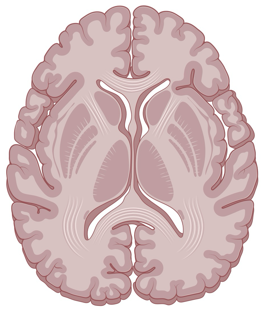
Glioma symptoms depend heavily on where in the brain the tumor is located. Select a brain region below to see the symptoms typically associated with tumors there.

Axial brain slice showing approximate lobe positions Frontal Temporal Parietal Occipital

Axial slice · approximate lobe positions shown for orientation

Symptoms That Occur Regardless of Location

Some symptoms are common to gliomas anywhere in the brain, because they result from the tumor's overall presence, associated swelling, or pressure inside the skull, rather than from damage to one specific region.

Headaches Seizures Nausea & vomiting Fatigue Personality or mood changes New depression or anxiety Trouble thinking, learning, or remembering Dizziness or loss of balance

Understanding Tumor Grade

The World Health Organization (WHO) grades gliomas from 1 to 4 based on how the cells look under the microscope and their molecular features. Grade is one of the most important factors shaping your treatment plan and outlook.

What Determines My Treatment Plan?

No two gliomas are treated identically. Several factors, some about the tumor itself and some about you, shape what surgery, radiation, and chemotherapy plan makes sense.

👆

Select a category above, then tap any factor to read more.

My Treatment Journey — Step by Step

Glioma treatment is a multi-stage process, not a single operation. Select each step to learn what happens and why.

👆 Select a step to learn what happens — and why.

What Outcomes Can Patients Expect?

Outcomes vary enormously by tumor grade, molecular profile, extent of resection, and your age and overall health. The figures below are population averages, not predictions for any individual.

10–15 years
Grade 2 (Low-Grade) Median Survival
With surgery and, when appropriate, radiation or chemotherapy. Many grade 2 tumors eventually recur or progress to a higher grade over time.
2–7 years
Grade 3 Median Survival
A wide range reflecting differences in molecular subtype; tumors with IDH mutation and 1p/19q co-deletion tend toward the more favorable end.
12–15 months
Glioblastoma (Grade 4) Median Survival
With standard treatment (surgery, radiation, and temozolomide chemotherapy). Roughly 5–7% of patients survive 5 years or more; younger patients and those with MGMT-methylated tumors tend to do better.

Why Extent of Resection Matters

Across many studies, removing a greater proportion of the visible tumor, when this can be done safely, is consistently associated with longer survival and better seizure control. This is the reasoning behind tools like image guidance, 5-ALA fluorescence, and awake mapping: they let the surgical team push the extent of resection as far as possible without crossing into tissue whose function cannot be restored.

Risks and Side Effects

Surgery: risks depend heavily on tumor location and include new or worsened weakness, numbness, speech difficulty, or vision changes; infection; bleeding; and seizures. Awake mapping is specifically used to reduce the risk of permanent deficits when a tumor sits near critical pathways.

Radiation: fatigue, hair loss at the treatment site, skin irritation, and, over months to years, changes in memory or concentration. Radiation necrosis, tissue injury that can mimic tumor regrowth on imaging, is an uncommon but recognized delayed effect.

Chemotherapy: fatigue, nausea, and lowered blood cell counts (raising infection and bleeding risk) are the most common effects of temozolomide and related drugs. Tumor Treating Fields (Optune), an alternative or add-on to chemotherapy, most commonly causes scalp irritation from the adhesive arrays.

Glioma Care at Brown

Brown Neurosurgery's Comprehensive Brain Tumor Center brings surgery, neuro-oncology, radiation oncology, neuropathology, and neuroradiology together for every glioma patient, rather than treating each specialty in isolation.

Every glioma case is reviewed by a multidisciplinary team, brain tumor-focused neurosurgeons, neuro-oncologists, radiation oncologists, neuroradiologists, and neuropathologists, so that the surgical plan, the pathology and molecular workup, and any radiation or chemotherapy plan are built together rather than handed off in sequence. Because gliomas are located deep within the brain and grow diffusely into surrounding tissue, complete surgical removal is not always possible; our team's first step is a maximal safe resection, followed by whichever combination of chemotherapy, radiation, and other therapies your specific tumor calls for.

Our program maintains a dedicated glioma research laboratory at Rhode Island Hospital, active clinical trials for new glioma treatments, and specific research programs in brain tumor immunotherapy and in Tumor Treating Fields. Consideration of alternating electrical fields (Optune) and clinical trials, including approaches involving immune modulation, are discussed with each eligible patient as part of building a full treatment plan, not offered only as a last resort.

Technology & Programs
Comprehensive Brain Tumor CenterMultidisciplinary review of every case among neurosurgery, neuro-oncology, radiation oncology, neuroradiology, and neuropathology.
Gamma Knife & CyberKnife RadiosurgeryFocused radiation delivery for residual, recurrent, or surgically inaccessible tumor.
TrueBeam RadiotherapyPrecision fractionated radiation therapy for the standard 6-week treatment course.
Brain Tumor Immunotherapy ProgramResearch and clinical trials exploring immune-based approaches to glioma treatment.
Dedicated Glioma Research LaboratoryRhode Island Hospital-based laboratory studying glioma biology and Tumor Treating Fields, working toward new therapies.

Common Questions

Will I be awake during my surgery?
Not always. Whether any part of your surgery is done awake depends on where the tumor is. If it sits near brain regions that control speech, movement, or other functions we cannot reliably map any other way, the surgical team will keep you awake and lightly sedated for a portion of the operation so those functions can be tested directly. If the tumor is farther from these areas, the entire procedure can be done under general anesthesia.
Can my whole tumor be removed?
Sometimes, but not always, and that is expected rather than a sign that something went wrong. Because gliomas grow diffusely into surrounding brain tissue without a clear edge, the surgical goal is usually maximal safe resection: removing as much tumor as possible without damaging brain function. Even when some tumor remains, a large resection still meaningfully improves survival and symptom control, and any remaining tumor is typically addressed with radiation or chemotherapy.
What is 5-ALA, and how does it help my surgeon?
5-ALA is a drug taken by mouth several hours before surgery. It is absorbed selectively by certain glioma cells and makes them fluoresce pink under a special surgical microscope light, letting the surgeon distinguish tumor from normal brain tissue in real time and remove more of the tumor's diffuse margin than could be seen with the naked eye alone.
What happens to the tumor tissue after surgery?
The tissue is sent to neuropathology, where it is examined under the microscope and tested for specific gene mutations (including IDH, MGMT, and 1p/19q status). This typically takes one to two weeks and provides the final diagnosis, grade, and molecular profile that determines your radiation and chemotherapy plan.
Will I need radiation and chemotherapy after surgery?
Most grade 2 through 4 gliomas are treated with radiation, chemotherapy, or both after surgery, since microscopic tumor cells extend beyond what surgery alone can remove. Some grade 1 and select grade 2 tumors, especially those completely removed, may be followed with imaging alone. Your neuro-oncology team will recommend a plan based on your tumor's grade and molecular profile.
What is Tumor Treating Fields (Optune), and will I need it?
Optune is a wearable device that delivers alternating electrical fields through adhesive scalp arrays to slow tumor cell division. It is most often considered for newly diagnosed or recurrent glioblastoma, typically alongside chemotherapy, and requires shaving the scalp and wearing the device most hours of the day. Not every patient is a candidate or wants to use it; your team will discuss whether it fits your specific situation.
Can a glioma come back after treatment?
Yes, recurrence is common, particularly for grade 3 and 4 gliomas, and typically occurs near the site of the original tumor. This is why regular surveillance MRI continues for years after treatment. If a tumor recurs, options can include additional surgery, radiation, a change in chemotherapy, or a clinical trial, depending on what you have already received.
What is the difference between a low-grade and a high-grade glioma?
Grade 1 and 2 tumors ("low-grade") grow slowly, and some can be cured or controlled for many years, though grade 2 tumors can eventually transform into a higher grade. Grade 3 and 4 tumors ("high-grade") grow and spread faster, are harder to remove completely, and require more intensive combined treatment. Grade is determined by how the cells look under the microscope and specific molecular markers, not by tumor size alone.
Will I be able to drive after my diagnosis?
If you have had a seizure or are on anti-seizure medication, driving is typically restricted for a period defined by your state's regulations, often at least several months seizure-free. Your care team will review your specific situation and let you know when it is safe and legal to resume driving.

Meet the Team

Glioma care at Brown draws on neurosurgery, neuro-oncology, and radiation oncology working as one team from diagnosis through long-term follow-up.

Clark C. Chen, MD, PhD
Director, Brain Tumor Program
Leads surgical care for glioma, integrating advanced imaging and molecular profiling into every case reviewed at the Comprehensive Brain Tumor Center.
Heinrich Elinzano, MD
Neuro-Oncology Medical Lead
Directs chemotherapy and systemic treatment planning, and co-leads the Brain Cancer Committee at Brown University Health Cancer Institute.
Jaroslaw T. Hepel, MD
Director, Stereotactic Radiosurgery, Rhode Island Hospital
Oversees radiation therapy planning, including fractionated radiotherapy and radiosurgery for residual or recurrent tumor.

See full profiles and the complete Comprehensive Brain Tumor Center team on the Brown Neurosurgery website.

What Is Glioma? Symptoms Tumor Grades Treatment Planning My Journey Outcomes